
Contact Info
Adam P. Hitchcock
Canada Research Chair
in Materials Research
CLS-CCRS
B.I.M.R
McMaster University
Hamilton, ON
Canada L8S 4M1
V: +1 905 525-9140
x24729
F: +1 905 521-2773
E: aph@mcmaster.ca
U: unicorn.mcmaster.ca
__________
Research
Group
Opportunities
Publications
Links
_____________

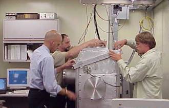
WHO: Harald Ade, David Kilcoyne (NCSU), Tony Warwick Keith Franck, Erik Anderson, Bruce Harteneck (LBNL), Adam Hitchcock, Tolek Tyliszczak, Peter Hitchcock (McMaster)
WHERE: Advanced Light
Source BL
5.3.2 Scanning Transmission X-ray Microscope (STXM)
WHEN: May 2001 to Jan 2002
POSTED: 23 Apr 2002
WHAT: Over the past three years a qualitatively new design
of scanning transmission X-ray microscope has been conceived, built
and commissioned at the Advanced Light Source. A dramatic improvement
in performance has been achieved by introducing laser interferometry
to control the relative spacing of microscope elements in order to achieve
a mechanical precision consistent with the optical resolution, currently
about 35 nm.
The interferometer based system is a great advance over previous systems
(old BL7.0 STXM at ALS; NSLS, X1a, Stony Brook). In the latter, one
could image at a fixed photon energy with a spatial resolution close
to the diffraction limited zone plate performance, but the spatial resolution
was effectively degraded for chemical analysis applications because
it was not possible to change photon energy while maintaining the focused
X-ray spot on the same place on a sample, or to image at successive
energies over the same x-y frame. The reason is the strongly chromatic
nature of zone plates. Over a typical C 1s NEXAFS scan from 280 to 310
eV the focal length changes by up to 300 mm, requiring displacement
of the zone plate along the X-ray beam by this amount while preserving
the lateral position of the zone plate relative to the sample. Mechanical
systems to achieve linearity of 1 in 104, with adjustment of the motion
axis to the X-ray axis to a similar precision are exceptional, and indeed,
the experience at both the ALS and NSLS microscopes has been that spatial
resolution in point spectral mode is rarely better than 200 nm and that
one needs to use software to correct for image-to-image misalignment,
with concomitant residual resolution degradation. 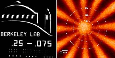
The solution that has been developed at the ALS 5.3.2 STXM is to introduce
a 2-d laser interferometer which is used as part of the feedback control
loop for the fast piezo stage used to position and scan the sample when
making images. This system provides stability of better than 10 nm with
response times up to 100 Hz and thus it not only eliminates energy-to-energy
image position errors, but also helps to desensitize the microscope
to vibrational or other environmental noise. With this mechanical/optical/control
system, accompanied by advanced control and acquisition software, the
5.3.2 STXM is able to achieve diffraction limited spatial resolution.
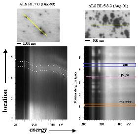
Fig. 1 shows images of Au test patterns in which periodic features with a half period of 30 nm could be observed with a zone plate that has a theoretical Rayleigh resolution of 45 nm. More significantly for analytical applications, the interferometry allows the 5.3.2 microscope to retain image registry at all photon energies. As an example of the level of positional stability achieved with the interferometry system, Fig. 2 compares linescan spectral images acquired at BL 5.3.2 on a polyurethane sample containing nanoscale filler particles (courtesy Ed Rightor, Dow Chemical) with a data set on a similar sample recorded over a similar spatial and photon energy range, acquired witha STXM without interferometric position control (the old ALS beamline 7.0 STXM). The 'image' is a plot of position along the line indicated in the image versus photon energy. Horizontal edges on the contrast features indicate exactly the same line is traced at each photon energy.
Clearly the ability to determine the NEXAFS spectra at high spatial
resolution is greatly enhanced by the improved performance of the 5.3.2
STXM.
WANT FURTHER INFORMATION ? : progress
report Jan-02 or CONTACT Adam Hitchcock
© 2002 A. Hitchcock / McMaster University
All Rights Reserved
Last updated by Christopher Amis on 04/23/2002
