Contact Info
Adam P. Hitchcock
Chemistry & ChemBio
B.I.M.R
McMaster University
Hamilton, ON
Canada L8S 4M1
V: +1 905 525-9140
x24749
F: +1 905 521-2773
E: aph@mcmaster.ca
U: unicorn.mcmaster.ca
__________
Research
Group
Opportunities
Publications
Links
_________

RECENT PUBLICATIONS:
Sep 2024 P.Ingino, H. Eraky,
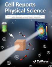
C. Zhang, A. P. Hitchcock and M. Obst, Soft X-ray spectromicroscopic
proof of a reversible oxidation/reduction
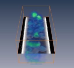
of individual microbial biofilm structures
using a novel microfluidic in situ electrochemical device,
Scientific Reports 14 (2024) 24009.
doi.org/10.1038/s41598-024-74768-9
Sep 2024 A.P. Hitchcock et al.,
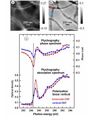
Comparison of Soft X-ray spectro-ptychography and Scanning Transmission
X-ray Microscopy,
J. Electron Spec. & Rela. Phen 276 (2024) 147487. doi.org/10.1016/j.elspec.2024.147487
Mar 2024 H. Eraky, J.J. Dynes and A. P. Hitchcock,
Mn 2p and O 1s X-ray absorption spectroscopy of manganese oxides,
J. Electron Spec. & Rela. Phen.274 (2024) 147452.
doi.org/10.1016/j.elspec.2024.147452
LATEST VERSION of aXis2000 - 01 Jan 2026
LATEST VERSION of soft X-ray Microscopy Bibliography - 09 Oct 2024
The Hitchcock group develops new instruments, methods and
applications of inner shell excitation spectroscopies and
microscopies for analysis of advanced materials. We are
particularly active in the application of X-ray microscopy to
energy materials [PEM fuel cells, CO2 reduction catalysts,
supercapacitors], polymers, nanostructures, biomaterials,
magnetotactic bacteria, environmental studies. A focus at present
is in situ and operando methods, such as
in situ flow electrochemistry, and tge investigation
of polymer electrolyte membrane fuel cell electrodes under
realistic (T,RH. We also use electron impact and synchrotron
methods to advance our understanding ot the fundamentals of
Inner shell excitation spectroscopy of gases, surfaces,
solids and liquids.
A common theme of all projects is the development of new
experimental techniques based on electron impact and X-ray
absorption phenomena, and optimization of their application to
a wide variety of chemical, physical and materials problems.
Collaborations with researchers in industry, government, and other
universities play a large role in our approach to promote interest
in, and practical uses for, inner-shell spectroscopies. Typically,
these collaborations combine the group's expertise in X-ray and
electron impact spectroscopic analysis with chemical, materials or
physicsl problems motivated by the collaborating group.
Our group has helped develop the most advanced analytical soft X-ray microscope in the world (a scanning transmission X-ray microscope at the Advanced Light Source in Berkeley, and lead the mplementation of STXMs at the Canadian Light Source (CLS) in Saskatoon). Over the past 5 years, in collaboration with instrument developers and scientists at the CLS, we have helped develop a new cryo-STXM with capabilities for cryo-spectro-tomography. We are helping to develop analysis software and applications of soft X-ray ptychography including the first demonstration of ptychography at the C 1s edge.
The CLS soft X-ray spectroscopy beamline (SM) features two scanning transmission X-ray microscopes (STXM) and an X-ray photoemission electron microscope (X-PEEM) on separate branch lines of a high performance plane grating monochromator beamline, illuminated by an elliptically polarized undulator. In October 2022, a new monochomator was installed, providing enhanced performance over the full 130 -3000 eV range, with a 10-fold improvement abouve 2000 eV, due to introduction of a wide-range, multilayer coating.
Please explore the pages of this web site. It contains highlights of our research, information about group members, opportunities for research positions, a list our publications (with links to pdfs), and links to tools we provide to others - aXis2000, BAN, the Core Excitation Bibiography and Database (~600 spectra of ~400 gas phase compounds!!), and a X-ray microscopy bibliography as well as links to related sites.
Enjoy your visit !
| |
|||||||
© 2054 A.P. Hitchcock / McMaster
University - All Rights Reserved
web page design by Christopher Amis (2002). Last updated on 01 Jan 2026
(aph)


