Contact Info
Adam P. Hitchcock
Canada Research Chair
in Materials Research
CLS-CCRS
B.I.M.R
McMaster University
Hamilton, ON
Canada L8S 4M1
V: +1 905 525-9140
x24729
F: +1 905 521-2773
E: aph@mcmaster.ca
U: unicorn.mcmaster.ca
__________
Research
Group
Opportunities
Publications
Links
_____________

Ptychography of a Magnetotactic Bacterium
WHO: Xiaohui Zhu, Adam Hitchcock (Chemistry &Chemical
Biology, BIMR, McMaster University )
)
Dennis
Bazylinski (UNLV), Ulysses Lins (UFRJ, Brasil)
Tolek
Tyliszczak, David Shapiro (ALS, LBNL)
WHERE: Advanced Light Source Scanning Transmission
X-ray Microscope (STXM11.0.2)
&
NanoSurveyor-1 (BL5.3.2.1)
WHEN: 2015-11 to 2016-09
POSTED: 29 Mar 2017, update 26-Jun-2017
WHAT: Spectro-ptychography X-ray absorption and X-ray Magnetic Cicular Dichroism at sub-10 nm resolution
Magnetotactic bacteria (MTB) synthesize chains of magnetic nanocrystals
(magnetosomes) that interact with the Earth's magnetic field like
an inner compass needle, simplifying their search for their optimum
environment at the oxic-anoxic interface.
How do these magnetosomes form? Some studies suggest that
hematite or amorphous ferrihydrite act as precursors, while others
suggest that they are formed directly from solution-phase Fe(II)
and Fe(III). Thus, identifying the chemical states of precursors
would be a good way to differentiate among competing models. Ptychography
is a coherent diffractive imaging technique with high resolution
(7 nm in this work) and excellent chemical-state sensitivity.
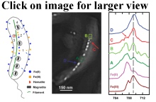
At ALS beamlines 5.3.2.1 and 11.0.2, we have measured ptychographic absorption and phase spectra of magnetosomes (~40-50 nm in size) inside a single cell of a marine MTB (Magnetovibrio blakemorei strain MV-1). The spectra of mature magnetosomes, immature magnetosomes, precursor regions, and the gaps between magnetosome chains differ, indicaitng that different iron species can coexist in a single cell.
From these results we proposed a model in which soluble Fe(II) is taken up from the environment, is partially oxidized to Fe(III), which in turn is then oxidized and transformed into hematite and ultimately into magnetite. In addition, ptychography ay BL 11.0.2 was used to measure the x-ray magnetic circular dichroism (XMCD) phase and absorption spectra of both extracellular and intracellular magnetosomes. Fe 2p XMCD probes the magnetism of magnetitein a site-specific manner. These experiments demonstrate that ptychography, which combines high spatial resolution, high-sensitivity chemical speciation, and site-specific magnetic information, offers a powerful probe for biomineralization studies.
OTHER INFORMATION:
Publication: X.H. Zhu, A.P. Hitchcock, D.A. Bazylinski, P. Denes, J. Joseph, U. Lins, S. Marchesini, H.-W. Shiu, T. Tyliszczak and D.A. Shapiro, Measuring spectroscopy and magnetism of extracted and intracellular magnetosomes using soft X-ray ptychography, Proc. Nat. Acad. Sci. 113 (51) (2016) E8219-E8227 doi:10.1073/pnas.1610260114
ALS Science Brief (published 14 Mar 2017) https://als.lbl.gov/ptychography-bacteriums-inner-compass/
© 2017 A.P. Hitchcock / McMaster University
- All Rights Reserved
web site design by Christopher Amis (2002). Last updated on 26 Jun 2017
(aph)

